| dc.creator | Sevick-Muraca, Eva | |
| dc.creator | Troy, Tamara L. | |
| dc.creator | Reynolds, Jeffery S. | |
| dc.date.accessioned | 2019-06-17T17:02:58Z | |
| dc.date.available | 2019-06-17T17:02:58Z | |
| dc.date.issued | 2008-02-05 | |
| dc.identifier.uri | https://hdl.handle.net/1969.1/176871 | |
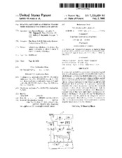
| dc.description.abstract | A system and method for non-invasive biomedical optical imaging and spectroscopy with low-level light is described. The technique includes a modulated light source coupled to tissue to introduce excitation light. Fluorescent light emitted in response to the excitation light is detected with a sensor. The AC intensity and phase of the excitation and detected fluorescent light is provided to a processor operatively coupled to the sensor. A processor employs the measured emission kinetics of excitation and fluorescent light to “map” the spatial variation of one or more fluorescence characteristics of the tissue and generate a corresponding image of the tissue via an output device. The fluorescence characteristic may be provided by exogenous contrast agents, endogenous fluorophores, or both. A technique to select or design an exogenous fluorescent contrast agent to improve image contrast is also disclosed. | en |
| dc.language | eng | |
| dc.publisher | United States. Patent and Trademark Office | |
| dc.rights | Public Domain (No copyright - United States) | en |
| dc.rights.uri | http://rightsstatements.org/vocab/NoC-US/1.0/ | |
| dc.title | Imaging of light scattering tissues with fluorescent contrast agents | en |
| dc.type | Utility patent | en |
| dc.format.digitalOrigin | reformatted digital | en |
| dc.description.country | US | |
| dc.contributor.assignee | The Texas A & M University System | |
| dc.identifier.patentapplicationnumber | 09/870144 | |
| dc.subject.uspcprimary | 600/473 | |
| dc.subject.uspcother | 600/431 | |
| dc.subject.uspcother | 600/476 | |
| dc.date.filed | 2001-05-30 | |
| dc.publisher.digital | Texas A&M University. Libraries | |
| dc.subject.cpcprimary | A61B 5/0059 | |
| dc.subject.cpcprimary | A61B 5/415 | |
| dc.subject.cpcprimary | A61B 5/418 | |